Treated conditions
Home » Areas of expertise » Disc Herniation
Treated conditions
Disc Herniation
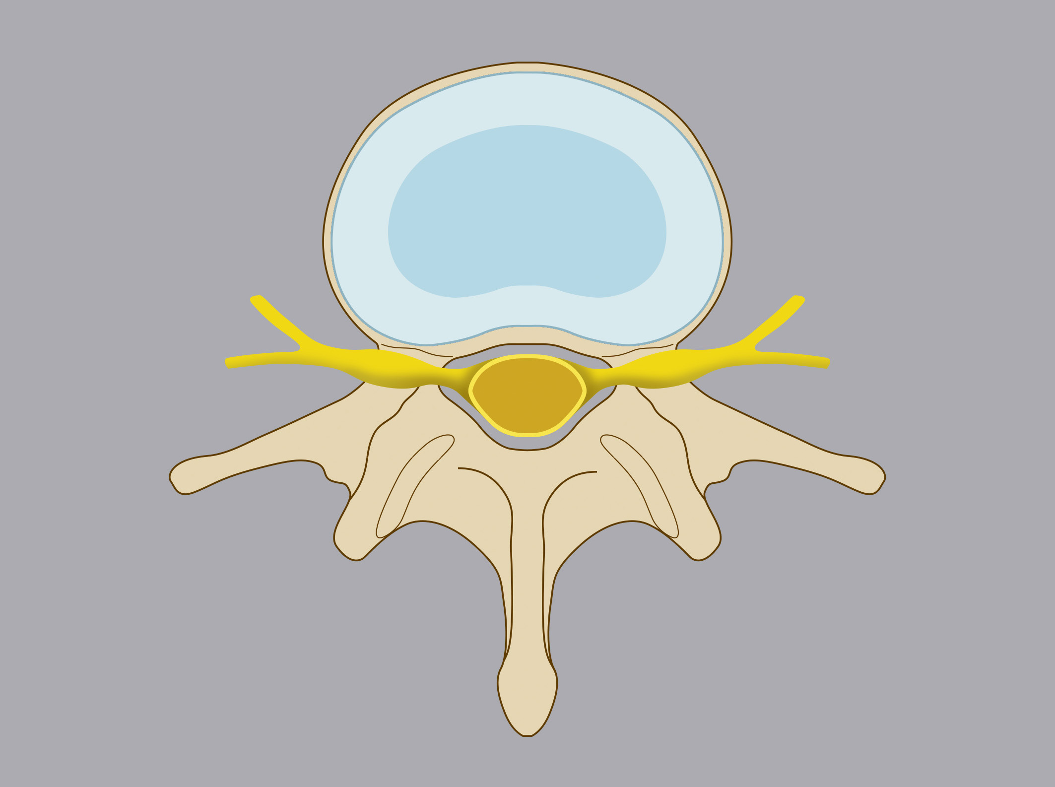
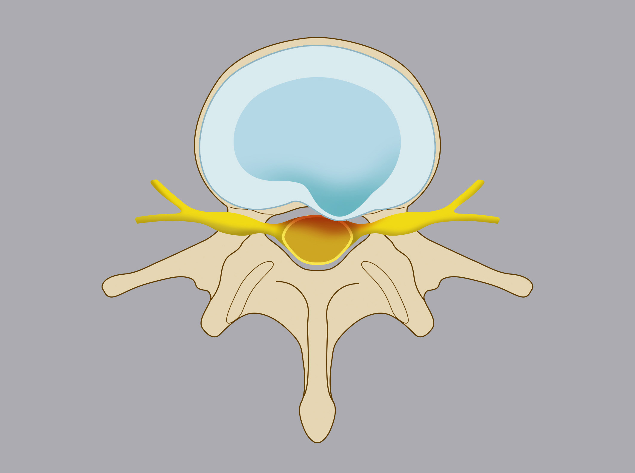
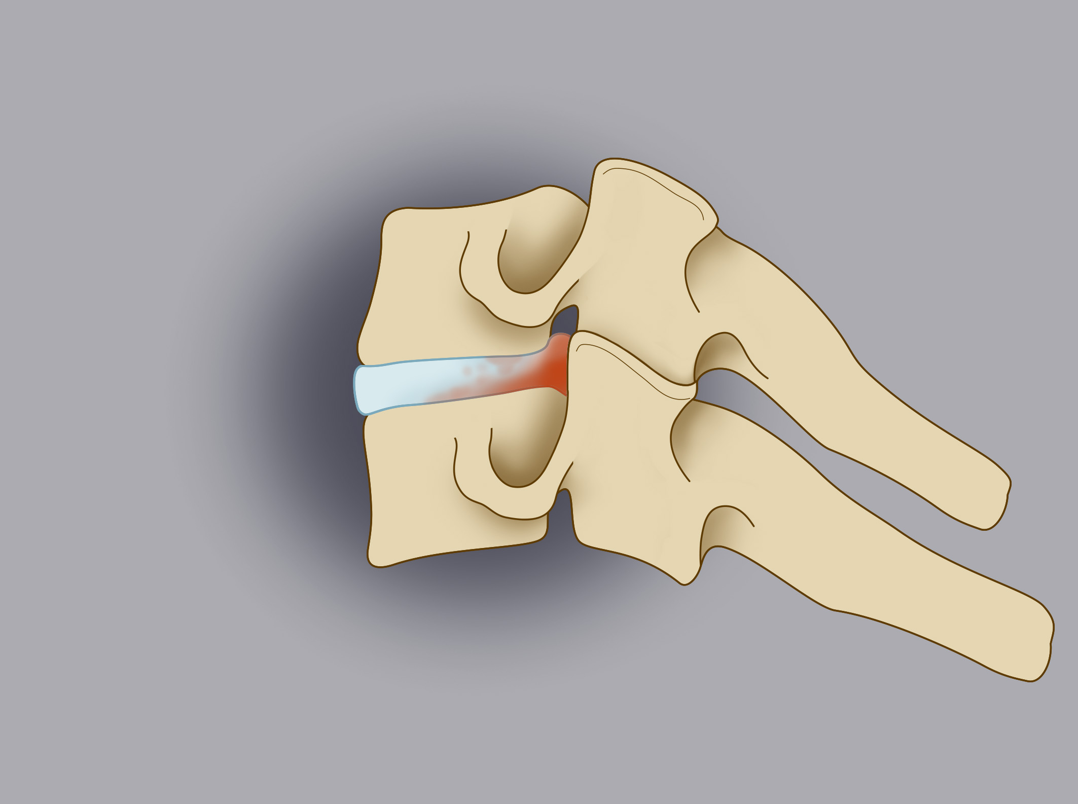
Intervertebral discs are fibrocartilaginous cushions between vertebrae, containing a central nucleus pulposus (90% water) and a peripheral annulus fibrosus. Degeneration due to prolonged load, poor posture, and physical strain can weaken the annulus fibrosus, leading to herniation. Herniated nucleus pulposus compresses spinal cord and/or nerve roots, causing symptoms.
Classification
Based on location:
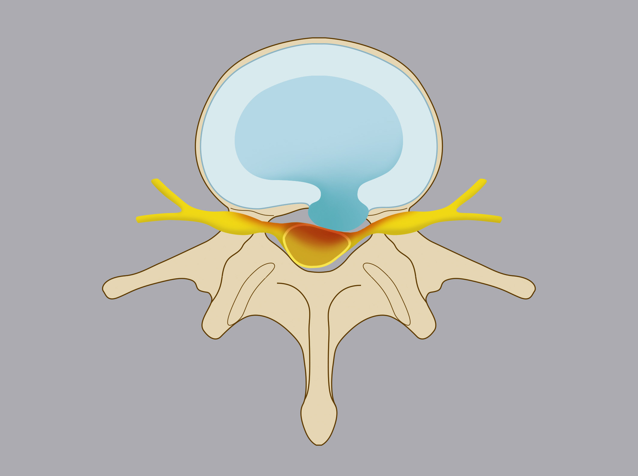
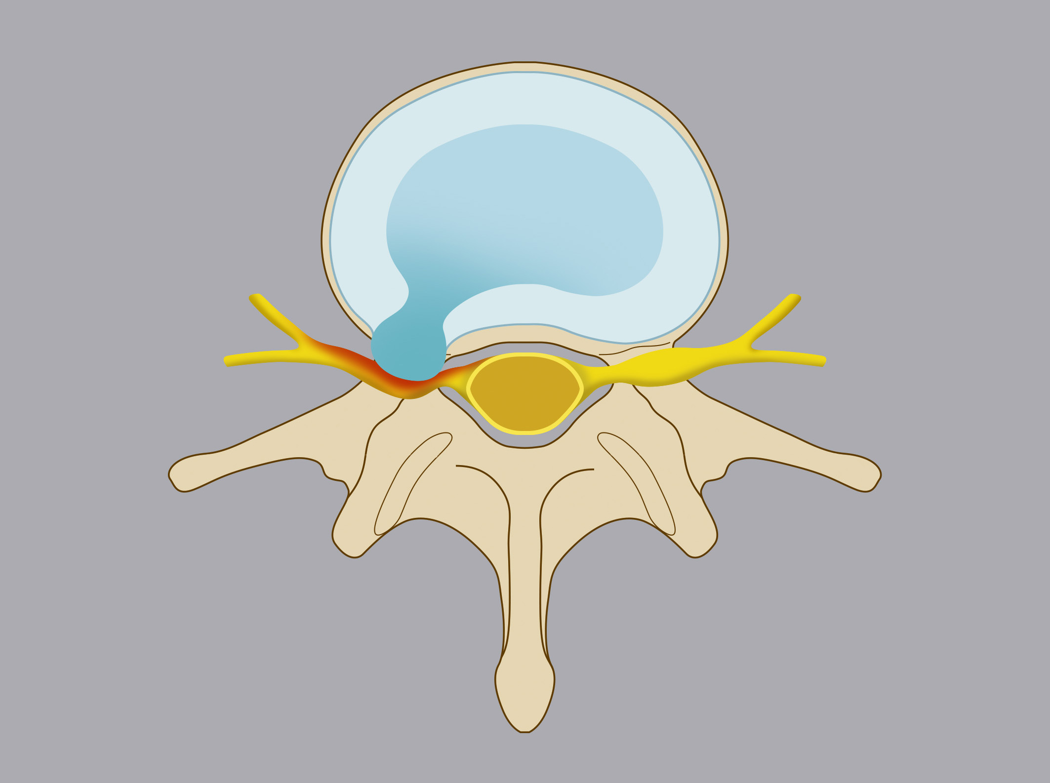
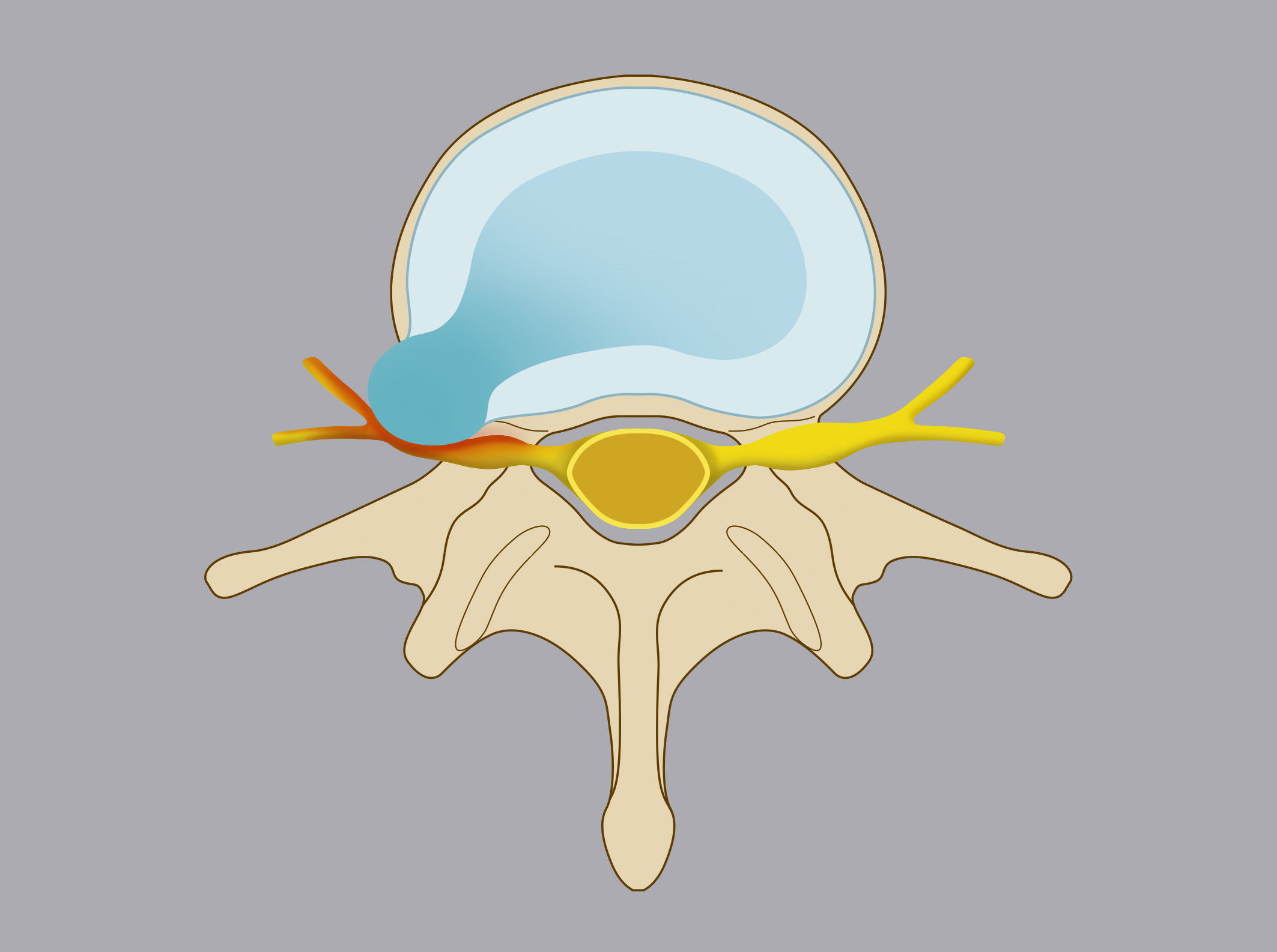
- Intracanal herniation: nucleus pulposus projects into the spinal canal
- Intraforaminal herniation: nucleus pulposus migrates into the neural foramen
- Extraforaminal herniation: nucleus pulposus migrates laterally to the neural foramen
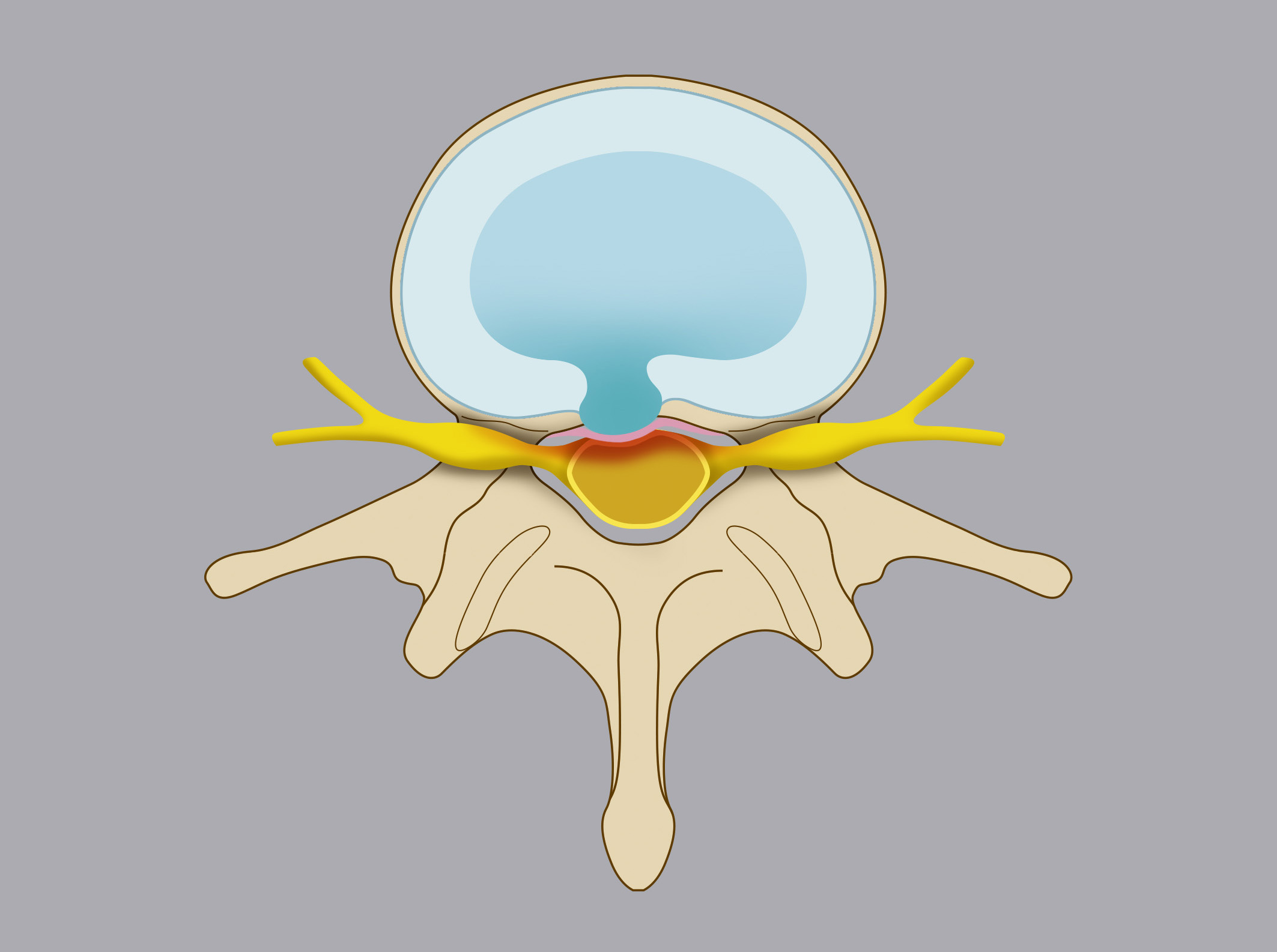
Based on nucleus pulposus protrusion:
- Disc protrusion: annulus fibrosus deforms without rupture
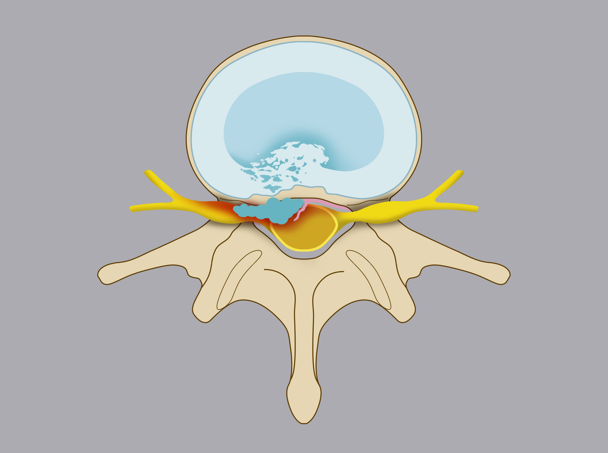
- Contained herniation: nucleus pulposus protrudes into the spinal canal but is contained by the posterior longitudinal ligament
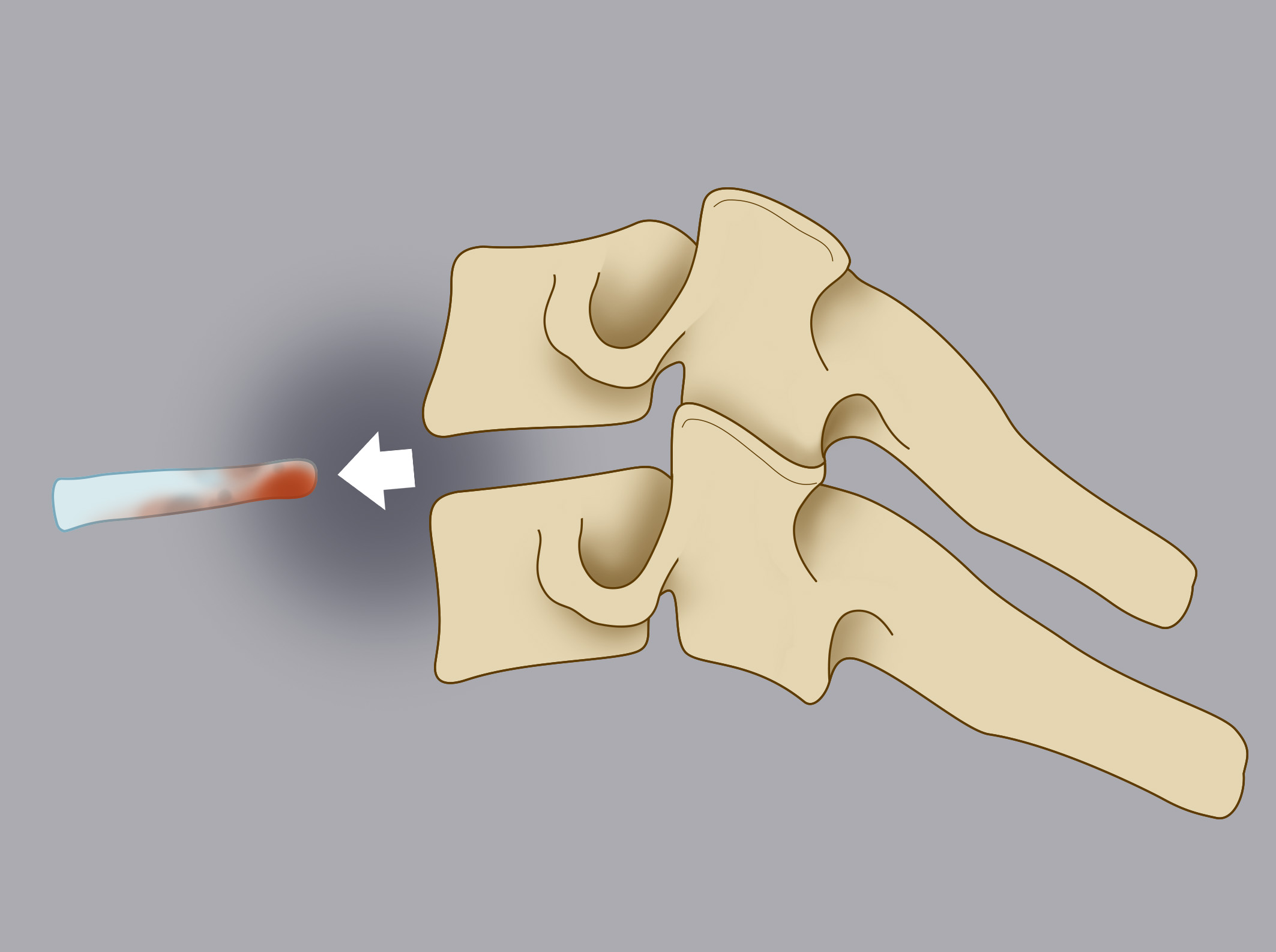
- Extruded herniation: nucleus pulposus ruptures through the annulus fibrosus and posterior longitudinal ligament
Causes
Excessive spinal load from physical labor, trauma, sports, sedentary lifestyle, obesity, genetic predisposition, and smoking.
Symptoms
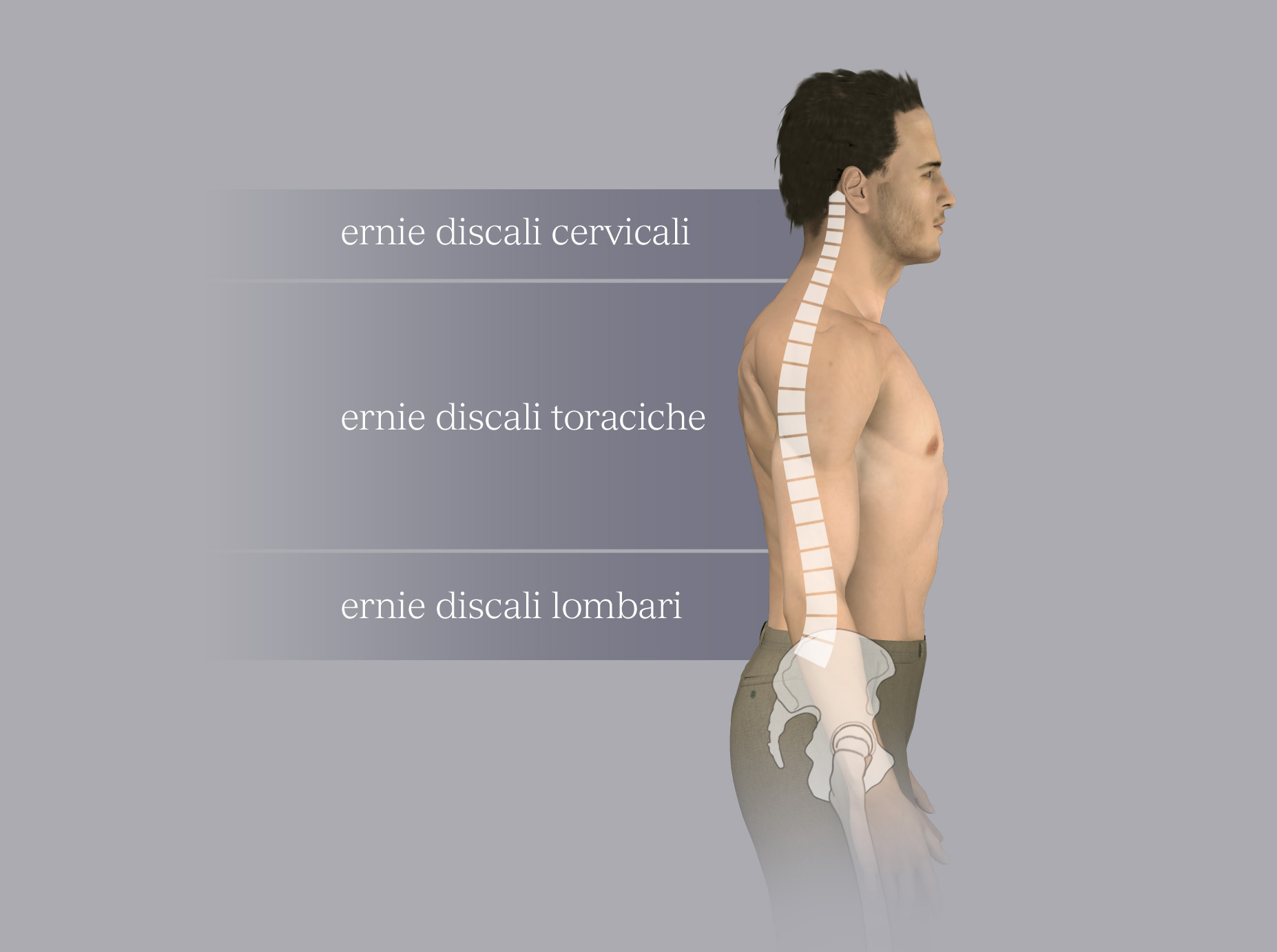
Cervical Disc Herniation
- Spinal cord compression: neck pain, upper/lower limb weakness, gait difficulty, urinary retention, constipation
- Nerve root compression: neck to arm pain, arm/hand numbness, muscle weakness
Thoracic Disc Herniation
- Spinal cord compression: back pain, lower limb weakness, gait difficulty, urinary retention, constipation, reduced sensitivity below compression
- Nerve root compression: back pain, intercostal pain, intercostal numbness
Lumbar Disc Herniation
- Nerve root compression: low back pain radiating to the leg, leg/foot numbness, muscle weakness
Diagnosis
MRI and CT scans confirm diagnosis, and electromyography (EMG) identifies affected nerve roots.
Treatment
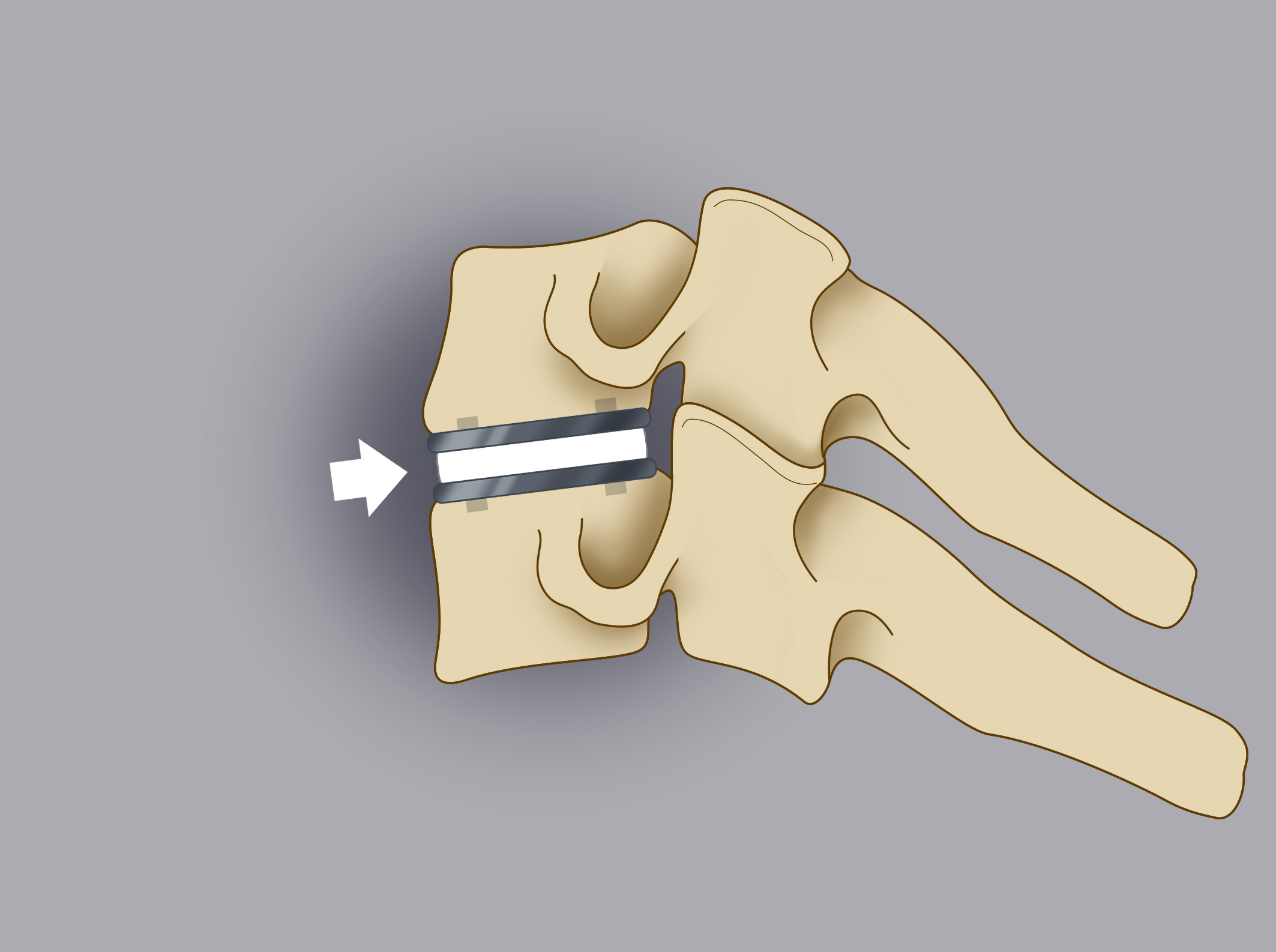
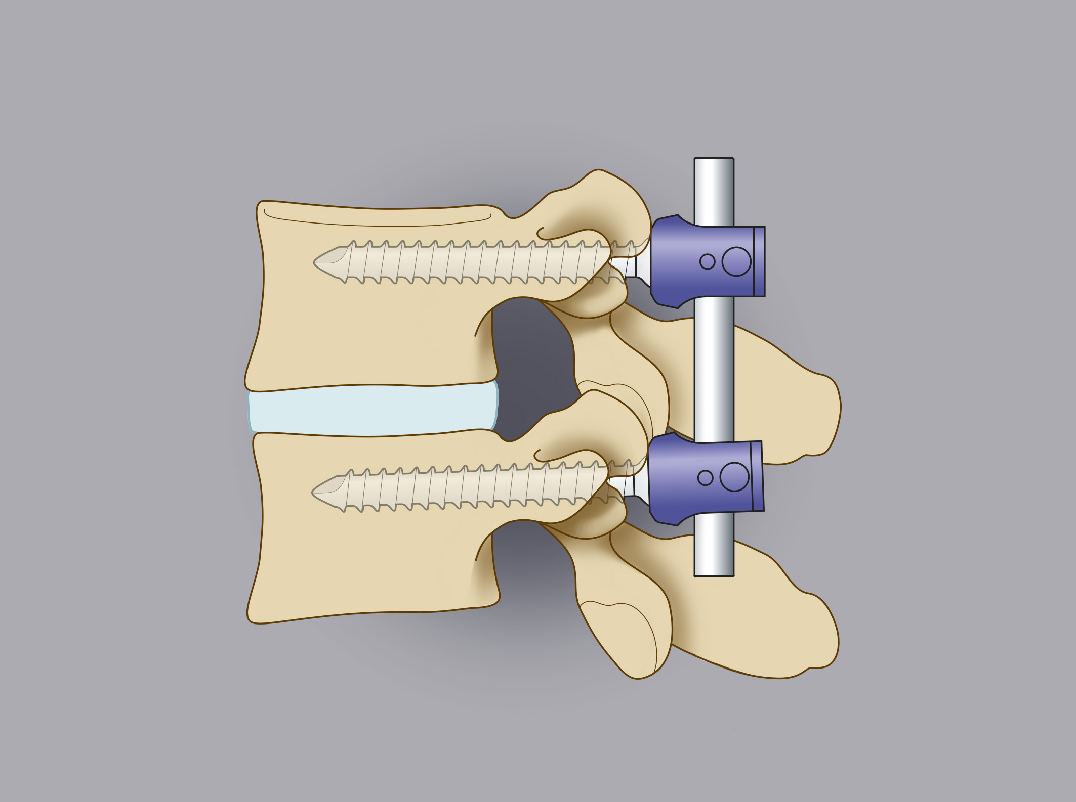
Most herniations resolve with conservative treatment (medication, physical therapy). Surgery (5-10% cases) is needed for motor deficits, severe pain, or incontinence, involving removal of herniated disc material to resolve nerve/spinal cord compression. Procedures include:
Cervical Herniation Surgery:
Anterior approach with intervertebral disc removal and replacement with a titanium/PEEK prosthesis.
Thoracic Herniation Surgery:
Lateral/antero-lateral approach, possibly involving partial rib removal or trans-thoracic approach with thoracic surgeons.
Lumbar Herniation Surgery:
Posterior approach with small incision for herniated fragment removal.